Meet the BCL-2 Family

Video originally posted by Genentech. From their site:

“Apoptosis is often evaded in cancer cells via overexpression of anti-apoptotic Bcl-2 family proteins and dysregulation of pro-apoptotic proteins. The Bcl-2 family members bind pro-apoptotic proteins to prevent apoptosis mediated by the intrinsic apoptotic pathway.

Bcl-2 is overexpressed in several hematologic malignancies, including non-Hodgkin’s lymphoma. Preclinical studies demonstrate that Bcl-2 acts as a key regulator of the intrinsic apoptotic signaling pathway by sequestering and neutralizing pro-apoptotic molecules, such as Bax.7 Thus, the anti-apoptotic protein promotes B-cell survival by inhibiting apoptosis, which may result in oncogenic chemotherapy resistance in hematologic malignancies”.

This cool image is also Genentech’s.

Impact of bone marrow stromal cells on Bcl-2 family members in chronic lymphocytic leukemia

The BCL-2 Family Reunion

Bodyguards and assassins: Bcl-2 family proteins and apoptosis control in chronic lymphocytic leukaemia

A new face of BCL-2 inhibition in CLL – inhibiting BCL-2 can promote cell death by perturbing calcium signaling!

“Zhong et al focus on a different facet of BCL-2, the BH4 domain that is involved in the interaction with IP3R. Using an oligopeptide derived from a site on IP3R found to be involved in binding BCL-2, the authors had previously demonstrated the ability to disrupt the BCL-2:IP3R complex and alter calcium signaling. This current report is noteworthy in two ways: first, it reports a modification of the peptide that increased cytoplasmic calcium concentrations; and second, it finds that CLL cells are selectively susceptible to death induced by the calcium signaling…”

Shikonin – another natural mitocan

“Shikonin, a natural naphthoquinone, was used in traditional Chinese medicine for the treatment of different inflammatory diseases and recent studies revealed the anticancer activities of shikonin. We found that shikonin has strong cytotoxic effects on 15 cancer cell lines, including multidrug-resistant cell lines. Transcriptome-wide mRNA expression studies showed that shikonin induced genetic pathways regulating cell cycle, mitochondrial function, levels of reactive oxygen species, and cytoskeletal formation. Taking advantage of the inherent fluorescence of shikonin, we analyzed its uptake and distribution in live cells with high spatial and temporal resolution using flow cytometry and confocal microscopy. Shikonin was specifically accumulated in the mitochondria, and this accumulation was associated with a shikonin-dependent deregulation of cellular Ca2+ and ROS levels. This deregulation led to a breakdown of the mitochondrial membrane potential, dysfunction of microtubules, cell-cycle arrest, and ultimately induction of apoptosis. Seeing as both the metabolism and the structure of mitochondria show marked differences between cancer cells and normal cells, shikonin is a promising candidate for the next generation of chemotherapy”.

Shikonin Directly Targets Mitochondria and Causes Mitochondrial Dysfunction in Cancer Cells

Shikonin circumvents cancer drug resistance by induction of a necroptotic death

shikonin

Metabolic Targets in the Crosshairs

“Mitochondria are emerging as idealized targets for anti-cancer drugs. One reason for this is that although these organelles are inherent to all cells, drugs are being developed that selectively target the mitochondria of malignant cells without adversely affecting those of normal cells. Such anticancer drugs destabilize cancer cell mitochondria and these compounds are referred to as mitocans, classified into several groups according to their mode of action and the location or nature of their specific drug targets. Many mitocans selectively interfere with the bioenergetic functions of cancer cell mitochondria, causing major disruptions often associated with ensuing overloads in ROS production leading to the induction of the intrinsic apoptotic pathway. This in-depth review describes the bases for the bioenergetic differences found between normal and cancer cell mitochondria, focusing on those essential changes occurring during malignancy that clinically may provide the most effective targets for mitocan development. A common theme emerging is that mitochondrially mediated ROS activation as a trigger for apoptosis offers a powerful basis for cancer therapy. Continued research in this area is likely to identify increasing numbers of novel agents that should prove highly effective against a variety of cancers with preferential toxicity towards malignant tissue, circumventing tumor resistance to the other more established therapeutic anti-cancer approaches”. Follow the links:

Targeting Cancer Metabolism: Dietary and Pharmacologic Interventions

Natural Compounds as Regulators of the Cancer Cell Metabolism

Bioenergetic pathways in tumor mitochondria as targets for cancer therapy and the importance of the ROS-induced apoptotic trigger

Choosing between glycolysis and oxidative phosphorylation: A tumor’s dilemma?

Targeting Cell Metabolism In Chronic Lymphocytic Leukaemia (CLL); A Viable Therapeutic Approach?

Stalling the Engine of Resistance: Targeting Cancer Metabolism to Overcome Therapeutic Resistance

Is Cancer a Metabolic Disease?

Cancer as a Metabolic Disease

Targeting mitochondria for cancer therapy

Mitochondrial permeability transition pore as a selective target for anti-cancer therapy

Mitochondrial uncoupling and the reprograming of intermediary metabolism in leukemia cells

Mitocans as Novel Agents for Anticancer Therapy: An Overview

Apoptosis: from biology to therapeutic targeting

Metabolic targets in the crosshairs

Metabolic targets in the cross hairs

DCA – turning on OxPhos

“Inhibition of mitochondrial pyruvate dehydrogenase kinase (PDK) by dichloroacetate may be exploited to reverse the abnormal metabolism of cancer cells from glycolysis to glucose oxidation. As PDK negatively regulates pyruvate dehydrogenase, dichloroacetate indirectly stimulates the pyruvate to acetyl-CoA conversion. Dichloroacetate has been shown to downregulate the aberrantly high mitochondrial membrane potential of cancer cells, increase mitochondrial ROS generation and activate K+ channels in malignant, but not in normal cells143. Dichloroacetate also upregulated the expression of the K+ channel Kv1.5, which is often underexpressed by tumour cells, through the transcription factor nuclear factor of activated T cells (NFAT1). Dichloroacetatenormalized mitochondrial functions were accompanied by reduced proliferation, increased apoptosis and suppressed tumour growth without apparent toxicity, suggesting that the mitochondria–NFAT–Kv axis and PDK represent promising anticancer drug targets”.

Sodium dichloroacetate exhibits anti-leukemic activity in B-chronic lymphocytic leukemia (B-CLL) and synergizes with the p53 activator Nutlin-3

The anti-leukemic activity of sodium dichloroacetate in p53mutated/null cells is mediated by a p53-independent ILF3/p21 pathway

Targeting mitochondria for cancer therapy

738px-Dichloroacetic-acid-2D-skeletal

Sodium dichloroacetate exhibits anti-leukemic activity in B-chronic lymphocytic leukemia (B-CLL) and synergizes with the p53 activator Nutlin-3

Sodium dichloroacetate selectively targets cells with defects in the mitochondrial ETC

Combination of Sulindac and Dichloroacetate Kills Cancer Cells via Oxidative Damage

CLL, BH3 Mimetics, and Apoptosis Round II – Meet Hyperforin

Time for another natural anti-cancer compound that works in a manner similar to gossypol; it up-regulates the pro-apoptotic BH3 protein Noxa. It comes from St. John’s Wort.

“We previously reported that hyperforin, a phloroglucinol purified from Hypericum perforatum, induces the mitochondrial pathway of caspase-dependent apoptosis in chronic lymphocytic leukemia (CLL) cells ex vivo, and that this effect is associated with upregulation of Noxa, a BH3-only protein of the Bcl-2 family. Here, we investigated the role of this upregulation in the pro-apoptotic activity of hyperforin in the cells of CLL patients and MEC-1 cell line. We found that the increase in Noxa expression is a time- and concentration-dependent effect of hyperforin occurring without change in Noxa mRNA levels. A post-translational regulation is suggested by the capacity of hyperforin to inhibit proteasome activity in CLL cells. Noxa silencing by siRNA reduces partially hyperforin-elicited apoptosis. Furthermore, treatment with hyperforin, which has no effect on the expression of the prosurvival protein Mcl-1, induces the interaction of Noxa with Mcl-1 and the dissociation of Mcl-1/Bak complex, revealing that upregulated Noxa displaces the proapoptotic protein Bak from Mcl-1. This effect is accompanied with Bak activation, known to allow the release of apoptogenic factors from mitochondria. Our data indicate that Noxa upregulation is one of the mechanisms by which hyperforin triggers CLL cell apoptosis. They also favor that new agents capable of mimicking specifically the BH3-only protein Noxa should be developed for apoptosis-based therapeutic strategy in CLL”.


Hyperforin structure

Hyperforin structure

Hyperforin induces apoptosis of chronic lymphocytic leukemia cells through upregulation of the BH3-only protein Noxa

Noxa upregulation is associated with apoptosis of chronic lymphocytic leukemia cells induced by hyperforin but not flavopiridol

Green Tea/Curcumin – a one-two punch for CLL

Once again we have natural compounds that have strong anti-cancer activity and effect multiple pathways. There’s quite a bit of overlap between these two, but also some antagonism as well, so if you decide to supplement these pay attention to the required dosing schedule.

EGCG curcumin

Curcumin Inhibits Pro-survival Pathways in CLL B-cells and has the Potential to Overcome Stromal Protection of CLL B-cells in Combination with EGCG

Turmeric and green tea: a recipe for B-Chronic Lymphocytic Leukemia

VEGF receptor phosphorylation status and apoptosis is modulated by a green tea component, epigallocatechin-3-gallate (EGCG), in B-cell chronic lymphocytic leukemia

Phase 2 Trial of Daily, Oral Polyphenon E in Patients with Asymptomatic, Rai Stage 0-II Chronic Lymphocytic Leukemia(CLL)

Orlistat – surprising OTC med targets lipid metabolism in CLL

“Constitutively activated pathways contribute to apoptosis resistance in chronic lymphocytic leukemia (CLL). Little is known about the metabolism of lipids and function of lipases in CLL cells. Performing gene expression profiling including B-cell receptor (BCR) stimulation of CLL cells in comparison to healthy donor CD5+ B cells, we found significant overexpression of lipases and phospholipases in CLL cells. In addition, we observed that the recently defined prognostic factor lipoprotein lipase (LPL) is induced by stimulation of BCR in CLL cells but not in CD5+ normal B cells. CLL cellular lysates exhibited significantly higher lipase activity compared to healthy donor controls. Incubation of primary CLL cells (n=26) with the lipase inhibitor orlistat resulted in induction of apoptosis, with a half-maximal dose (IC50) of 2.35 mum. In healthy B cells a significantly higher mean IC50 of 148.5 mum of orlistat was observed, while no apoptosis was induced in healthy peripheral blood mononuclear cells (PBMCs; P<0.001). Orlistat-mediated cytotoxicity was decreased by BCR stimulation. Finally, the cytotoxic effects of orlistat on primary CLL cells were enhanced by the simultaneous incubation with fludarabine (P=0.003). In summary, alterations of lipid metabolism are involved in CLL pathogenesis and might represent a novel therapeutic target in CLL”. Follow the link: Targeting lipid metabolism by the lipoprotein lipase inhibitor orlistat results in apoptosis of B-cell chronic lymphocytic leukemia cells

Orlistat_Structural_Formulae

Phenethyl Isothiocyanate (PEITC)

Watercress has it. So does cauliflower, cabbage, bok choy, broccoli, and brussels sprouts. Phenethyl Isothiocyanate (PEITC) is another powerful, natural anti-cancer compound. It works by manipulating redox status in the cell. Follow the links for some of the research on this powerful glutathione inhibitor.

Structure of PEITC

Structure of PEITC

Stromal control of cystine metabolism promotes cancer cell survival in chronic lymphocytic leukemia

Effective elimination of fludarabine-resistant CLL cells by PEITC through a redox-mediated mechanism

Overcoming resistance to histone deacetylase inhibitors in human leukemia with the redox modulating compound β-phenylethyl isothiocyanate

Inhibition of Mitochondrial Respiration and Rapid Depletion of Mitochondrial Glutathione by β-Phenethyl Isothiocyanate: Mechanisms for Anti-Leukemia Activity

Phenethyl Isothiocyanate (PEITC) Regulates Autophagy in Chronic Lymphocytic Leukemia

CLL, BH3 Mimetics, and Apoptosis

Update: Follow the link for a nice primer on apoptosis, BCL-2, and BH3 Mimetics.

Dr. Sharman’s CLL & Lymphoma Blog – What is BCL-2 and why should we inhibit it?

“Chronic lymphocytic leukemia (CLL) is characterized by the deregulated accumulation and persistence of B lymphocytes in the blood. Although the exact causes of CLL are unknown, the evasion of apoptosis through aberrant expression of BCL2-family proteins is a common feature. A class of compounds, termed BH3 mimetics, has been developed to directly inhibit BCL2 proteins and selectively kill tumor cells. To date, the most successful of these compounds are the BCL2/BCLXL inhibitors ABT-7372 and ABT-263 (navitoclax), as well as the BCL2-specific inhibitor ABT-199. Results from early clinical trials with navitoclax have demonstrated single-agent efficacy in patients with relapsed or refractory CLL.
However, there was heterogeneity in response rates between patients, and dose-limiting toxicities including thrombocytopenia and neutropenia which prevented further doseescalation.
In addition, CLL cells residing within various microenvironments (e.g. lymph nodes and bone marrow) are resistant to BCL2 inhibitors. This resistance results from the upregulation of additional BCL2-proteins, such as BCLXL, MCL1 and BFL1, the latter two of which are not inhibited by navitoclax, and therefore protect the leukemia cells from apoptosis. Additional drugs are needed to enhance the efficacy of navitoclax. Here, we demonstrate that gossypol overcomes stroma-mediated resistance to ABT-737 without enhancing the sensitivity of normal lymphocytes and platelets.
The BH3-only protein, NOXA, is a potent inhibitor of MCL1 and BFL1, but has recently been recognized to inhibit BCLXL with lower affinity. Therefore, compounds which induce NOXA may inhibit MCL1, BFL1 and BCLXL, thus overcoming resistance to navitoclax. We previously reported that six putative BH3 mimetics do not directly inhibit BCL2 in cells, but instead activate the integrated stress response and induce NOXA. Of these six compounds, gossypol has advanced into clinical trials in a racemically purified form (AT-101).10 We hypothesized that gossypol, through induction of NOXA, would sensitize CLL cells to ABT-737”. Link below:


Gossypol overcomes stroma-mediated resistance to the BCL2 inhibitor ABT-737 in chronic lymphocytic leukemia cells ex vivo

BH3 Mimetics – the road to apotosis

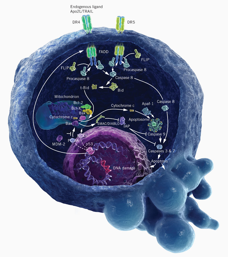
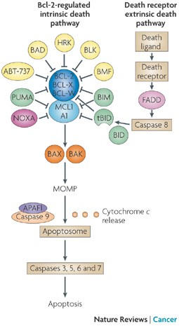
“In mammals, apoptosis occurs through the death receptor (extrinsic) or Bcl-2-regulated (intrinsic or mitochondrial) pathways. The latter is regulated by three subgroups of the Bcl-2 family: the pro-survival members, such as BCL-2 or MCL1, the pro-apoptotic BAX and Bcl-2 homologous killer (BAK) subgroup and the pro-apoptotic BCL-2 homology domain 3 (BH3)-only proteins, such as BIM (also known as BCL2L11) and PUMA (also known as BBC3). Apoptotic stimuli cause transcriptional and/or post-translational activation of specific BH3-only proteins, which then engage and sequester the pro-survival Bcl-2 family members, thereby liberating the downstream effectors, BAX and BAK, which elicit mitochondrial outer membrane permeabilization (MOMP) and unleash the caspase cascade, culminating in cell demolition. It has also been proposed that at least some BH3-only proteins, in particular BIM and BID, can directly activate BAX and BAK (not shown). Some BH3-only proteins (shown in green), such as BIM and PUMA, can bind and sequester all anti-apoptotic Bcl-2 family members with high affinity and are thus potent killers, whereas others (shown in yellow and dark pink), such as Bcl-2 antagonist of cell death (BAD) and NOXA (also known as PMAIP1), bind only certain anti-apoptotic members (BAD binds BCL-2, BCL-XL and BCL-W (dark blue), whereas NOXA binds only MCL1 and A1 (light blue)). Thus, the efficiency of cell killing is determined by the relative levels of pro- and anti-apoptotic members. ABT-737, a BH3-mimetic, has a similar binding profile to the BH3-only protein BAD. APAF1, apoptotic protease-activating factor 1; BMF, Bcl-2-modifying factor; HRK, activator of apoptosis harakiri; tBID, truncated BID.”


Links:

Multiple BH3 Mimetics Antagonize Antiapoptotic MCL1 Protein by Inducing the Endoplasmic Reticulum Stress Response and Up-regulating BH3-only Protein NOXA

Development of Noxa-like BH3 Mimetics for Apoptosis-Based Therapeutic Strategy in Chronic Lymphocytic Leukemia

Apoptosis: from biology to therapeutic targeting

Gossypol, a BH3 mimetic, induces apoptosis in chronic lymphocytic leukemia cells

Methyl Jasmonate – a natural anti-cancer compound with several modes of action

Methyl Jasmonate is a plant stress hormone that has significant anti-cancer properties. So how does MJ work? Let me count the ways. It arrests cell cycle, inhibiting cell growth and proliferation; causes cell death through the intrinsic/extrinsic pro-apoptotic, p53-independent apoptotic, and non-apoptotic (necrosis) pathways; detaches hexokinase from the voltage-dependent anion channel, dissociating glycolytic and mitochondrial functions, decreasing the mitochondrial membrane potential, favoring cytochrome c release and ATP depletion, activating pro-apoptotic and inactivating anti-apoptotic proteins; induces reactive oxygen species mediated responses; stimulates MAPK-stress signaling and redifferentiation in leukemia cells; inhibits overexpressed pro-inflammatory enzymes in cancer cells such as aldo-keto reductase 1 and 5-lipoxygenase; inhibits cell migration and shows antiangiogenic and anti-metastatic activities. The complete lack of toxicity to normal cells and the rapidity by which MJ causes damage to cancer cells, turns MJ into a promising anticancer agent that can be used alone or in combination with other agents.

Follow the links for the relevant research:

MJ modes of action

MJ modes of action

Methyl Jasmonate: Putative Mechanisms of Action on Cancer Cells Cycle, Metabolism, and Apoptosis

Jasmonates: Novel Anticancer Agents Acting Directly and Selectively on Human Cancer Cell Mitochondria

Methyl jasmonate abolishes the migration, invasion and angiogenesis of gastric cancer cells through down-regulation of matrix metalloproteinase 14

Methyl jasmonate Displays In Vitro and In Vivo Activity against Multiple Myeloma Cells

Jasmonates induce nonapoptotic death in high-resistance mutant p53-expressing B-lymphoma cells

Methyl jasmonate down-regulates survivin expression and sensitizes colon carcinoma cells towards TRAIL-induced cytotoxicity

Effects of natural and novel synthetic jasmonates in experimental metastatic melanoma